
Optical Coherence Tomography (OCT)
OCT provides 2D and 3D virtual cross-sectional images of an object in a non-invasive and non-contact way. It is suitable for the investigation of translucent and transparent materials (e.g. varnishes, glass, glazes). OCT is also used in a medical context, in Optician settings to create a detailed 3D image and assessment of eye health- thus proving the technique's safety and applicability to delicate and vulnerable structures!
ISAAC is equipped with a range of OCTs, the majority of which are developed in our Lab. These range in central wavelengths from the near- to the mid- infrared, offering a range of specifications from ultra-high resolution (~1 microns) to deep penetration into highly scattering materials. We also have a bespoke hybrid system which combines OCT and VIS/NIR Hyperspectral Imaging. Detailed specifications are given in the tables below.

Ultra High Resolution-OCT examining an easel painting from the National Gallery's collection- The Madonna and Child (NG929, after Raphael, probably before 1600)
© The National Gallery, London
Mounting of the probe head is flexible and various adaptations are possible. The speed of acquisition ranges from a few seconds to a few minutes for an image cube of a 1cm x 1cm area.
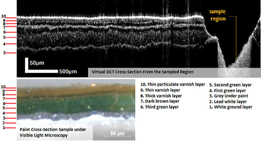
A sampled section of the above National Gallery painting: Top left: virtual OCT cross-section; Top right: 3D OCT image of the varnish and paint layers, directly imaged from the painting at a distance of a few cm. Bottom: paint cross section sample under Visible Light Microscopy
Applications OF OCT
-
high resolution and high contrast imaging of underdrawings
-
detection of delamination of internal layers, e.g. enamel
-
monitoring of varnish removal
-
monitoring of glass deterioration
-
non-contact examination of subsurface microstructure of intact objects
-
measuring the hydraulic conductivity of porous materials such as rock art
-
study of the manufacturing techniques of ancient artefacts etc.
OCT analysis of the Book of Hours of Isabella Stuart (Ms 62) at the Fitwilliam Museum, Cambridge


Colour image of enamel panel 1855,0305.2 from the British Museum Limoges enamel collection (white box indicates the OCT scanned area),along with the 3D video that shows the OCT cube. The subsurface structure, including the lower enamel layers and underdrawings, as well as the surface decoration can be seen.
ISAAC instrumentation
OCT System
Ultra-High Resolution OCT @810 nm
Telesto 2
@1310 nm
SWIR (Long Wavelength) OCT
@1960 nm
MWIR (Long Wavelength) OCT @3600 nm
Developer
ISAAC Lab
Thorlabs
ISAAC Lab
ISAAC Lab
Depth Penetration
Moderate
Moderate Deep
Deep
Deep
Depth Resolution (Air/Polymer)
1.8 µm / 1.2 µm
5.5 µm / 3.7 µm
9 µm / 6 µm
9 µm / 6 µm
Transverse Resolution
7 µm
15 µm – 20 µm
17 µm
27 µm
Working
Distance
64 mm
10 mm
40 mm
70 mm
Service
MOLAB/
FIXLAB
MOLAB/
FIXLAB
MOLAB/
FIXLAB
FIXLAB
Our in-house developed Hybrid OCT system combines an optical coherence tomography (OCT) system and a reflectance spectral imaging system into one instrument. OCT provides 2D and 3D virtual cross-sectional images in a non-invasive and non-contact way. VIS-NIR Spectral Imaging can provide identification of materials. The simultaneous use of the two analysis methods allows for a 1:1 spatial alignment between the spectral images and OCT 3D volumetric data sets, providing additional context on the layer structure.
ISAAC Hybrid OCT System:
Developer
OCT Depth Resolution (air/polymer)
OCT Transverse Resolution
Working Distance
Spectral Imaging Spectral Range
Spectral Imaging Spectral Resolution
Spectral Imaging Transverse Resolution
Hybrid OCT @ 1350 nm &
VIS/NIR Microscopic Spectral Imaging
ISAAC Lab
5 µm / 3.3 µm
10 µm
40 mm
415 nm – 845 nm
10 nm
5 µm
example Heritage science Projects
selected Publications
This is a selection of our OCT publications. Our full publication list can be found here.
Faluweki, M.K., Cheung, C.S., & Liang, H. 2023. Simultaneous Measurement of Refractive Index and Dispersion using Optical Coherence Tomography for Restoration of Transparent Works of Art. The European Physics Journal Plus, 138, 825. https://doi.org/10.1140/epjp/s13360-023-04458-4
Read, M., Cheung, C.S., Liang, H., Meek, A. and Korenberg, C. 2021. A Non-Invasive Investigation of Egyptian Faience Using Long Wavelength Optical Coherence Tomography (OCT) at 2µm. Studies in Conservation 67, 168-175. https://doi.org/10.1080/00393630.2020.1871208
Leona, M., Fukunaga, K., Liang, H., Baglioni, P., Festa, G. And Levchenko, V. 2021. From Physics to Art and Back. Nature Revews Physics 3, 681–684. https://doi.org/10.1038/S42254-021-00362-X
Read, M., Cheung, C.S., Ling, D., Korenberg, C., Meek, A., Kogou, S. and Liang, H., 2019. A Non-Invasive Investigation of Limoges Enamels using both Optical Coherence Tomography (OCT) and Spectral Imaging: A Pilot Study. In: H. Liang and R. Groves, eds., Optics for Arts, Architecture, and Archaeology Vii. SPIE Optical Metrology, Munich, Germany, 24-27 June 2019. SPIE Proceedings 11058, 1105803 https://doi.org/10.1117/12.2527092
Thickett, D., Cheung, C.S., Liang, H., Twydle, J., Maev, R.G. and Gavrilov, D., 2017. Using Non-Invasive Non-Destructive Techniques to Monitor Cultural Heritage Objects. Insight - Non-Destructive Testing and Condition Monitoring, 59 (5), pp. 230-234. http://doi.org/10.1784/insi.2017.59.5.230
Liang, H., Mari, M., Cheung, C.S., Kogou, S., Johnson, P. and Filippidis, G., 2017. Optical Coherence Tomography and Non-Linear Microscopy for Paintings – A Study of the Complementary Capabilities and Laser Degradation Effects. Optics Express, 25 (16), pp. 19640-19653. https://doi.org/10.1364/OE.25.019640
Cheung, C.S., Spring, M. and Liang, H., 2015. Ultra-High Resolution Fourier Domain Optical Coherence Tomography for Old Master Paintings. Optics Express, 23 (8), pp. 10145-10157. https://doi.org/10.1364/OE.23.010145
Cheung, C.S., Daniel, J.M.O., Tokurakawa, M., Clarkson, W.A. and Liang, H., 2015. High-Resolution Fourier Domain Optical Coherence Tomography in the 2 mm Wavelength Range using a Broadband Supercontinuum Source. Optics Express, 23 (3), pp. 1992-2001. https://doi.org/10.1364/OE.23.001992
Cheung, C.S., Daniel, J.M.O., Tokurakawa, M., Clarkson, W.A. and Liang, H., 2014. Optical Coherence Tomography in the 2-µm Wavelength Regime for Paint and Other High Opacity Materials. Optics Letters, 39 (22), pp. 6509-6512. https://doi.org/10.1364/OL.39.006509
Liang, H., Burgio, L., Bailey, K., Lucian, A., Bellesia, S., Cheung, C. and Brookes, C., 2014. Culture And Trade Through the Prism of Technical Art History: A Study of Chinese Export Paintings. Studies In Conservation, 59 (1). https://doi.org/10.1179/204705814X13975704318272
Cheung, C.S., Tokurakawa, M., Daniel, J., Clarkson, W.A. and Liang, H., 2013. Long Wavelength Optical Coherence Tomography for Painted Objects. Proceedings of SPIE, 8790, 87900j https://doi.org/10.1117/12.2021700
Bemand, E. and Liang, H., 2013. Optical Coherence Tomography for Vulnerability Assessment of Sandstone. Applied Optics, 52 (14), pp. 3387-3393. https://doi.org/10.1364/AO.52.003387
Liang, H., Lange, R., Peric, B. and Spring, M., 2013. Optimum Spectral Window for Imaging of Art with Optical Coherence Tomography. Applied Physics B: Lasers and Optics, 111 (4), pp. 589-602. https://doi.org/10.1007/s00340-013-5378-5
Cheung, C.S. and Liang, H., 2013. Ultra-High Resolution Fourier Domain Optical Coherence Tomography for Resolving Thin Layers in Painted Works of Art. Proceedings of SPIE, 8790, 87900m https://doi.org/10.1117/12.2020765
Bemand, E., Bencsik, M. and Liang, H., 2011. OCT and NMR for Non-Invasive In-Situ Monitoring of the Vulnerability of Rock Art Monuments. Proceedings of SPIE, 8084, 80840h https://doi.org/10.1117/12.890084
Lange, R., Liang, H., Howard, H. and Spooner, J., 2011. Optical Coherence Tomography and Spectral Imaging of a Wall Painting. SPIE Newsroom. https://irep.ntu.ac.uk/id/eprint/6042
Liang, H., Cid, M.G., Cucu, R.G., Dobre, G.M., Kudimov, B., Pedro, J., Saunders, D., Cupitt, J. and Podoleanu, A.G., 2005. Optical Coherence Tomography- A Non-Invasive Technique Applied to Conservation Of Paintings. Proceedings of SPIE 5857, 58570w https://doi.org/10.1117/12.612591
Liang, H., Cucu, R., Dobre, G.M., Jackson, D.A., Pedro, J., Pannell, C., Saunders, D. and Podoleanu, A.G., 2004. Application of OCT to Examination of Easel Paintings. Second European Workshop on Optical Fibre Sensors, 2004, 5502, pp. 378-381. https://doi.org/10.1117/12.566780